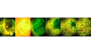
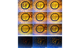
Winners: Earth Day Light Microscopy Imaging Competition
Lumencor’s Light Microscopy Imaging Competition celebrates Earth Day by highlighting our commitment to manufacturing sustainable microscope Light Engines that are bright, clean, and mercury-free.
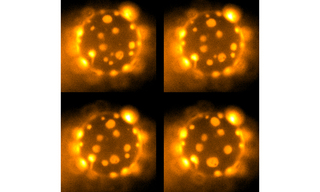
First Place Winner: Dr. Tejeshwar Rao
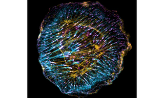
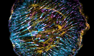
Second Place: Dr. Danaí Montalvan-Sorrosa
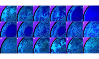
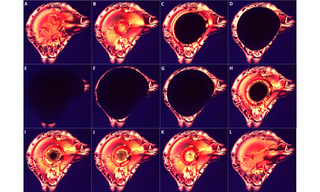
Third Place: Parthasarathy Raghuveer