BeRST-ing with Benefits: Transform Your Electrophysiology Studies with BeRST Voltage Sensitive Dye
Berkeley Red Sensor of Transmembrane Potential (BeRST) dye is a robust voltage-sensitive dye (VSD) designed for real-time monitoring of transmembrane action potential dynamics in excitable cells, such as cardiomyocytes and neurons. It is the ideal optical cellular physiology tool as it offers brightness, speed, and exceptional photostability [1]. Why should BeRST be the go-to dye for your optophysiology studies, as a direct analogue to what had been electrophysiology studies?
Multiplexing Compatibility
BeRST is a reliable membrane marker that has an excitation peak at 658 nm and an emission peak at 683 nm (Figure 1). It may be used alongside other common fluorescent markers at shorter wavelengths with minimal spectral overlap [1, 2]. Its near-infrared (nIR) spectral profile makes it perfect for multiplexed assays where multiple signals are tracked simultaneously with minimal interference or bleedthrough.Versatility for Cell Models and Applications
BeRST’s brightness makes it a suitable label for cell counting and morphometric analysis. It can be applied to both 2D and 3D cell models, offering flexibility for a wide range of experimental setups (Figure 2) [1, 2, 3, 4]. BeRST has been successfully used with channelrhodopsin (ChR2) to detect voltage changes and optogenetically control cell pacing in human-induced pluripotent stem cell-derived cardiomyocytes (hIPSC-CMs) [2, 5].Fast and Precise Fluorescence Response
BeRST fluorescence response occurs via photo-induced electron transfer (PeT), where changes in transmembrane voltage increase or decrease electron transfer efficiency and result in decreases or increases in fluorescence (Figure 3) [1]. This mechanism allows for faster and more precise detection of action potential changes compared to other voltage indicators, such as genetically encoded voltage indicators (GEVIs), which rely on slower protein conformational changes to track changes in voltage.
When BeRST is used as a VSD of choice with Lumencor’s VOLTA Scanner, remarkable 10 kHz sampling rates are realized, offering uniquely easy transmembrane dynamics reporting at high resolution with exceptional accuracy.
BeRST’s speed, sensitivity, and versatility make it the optimal tool for advancing your traditional electrophysiology studies with optical signals via optophysiology. Whether you are investigating cellular transmembrane dynamics or uncovering new insights into excitable cell behavior, BeRST provides the performance needed for precise, real-time monitoring.
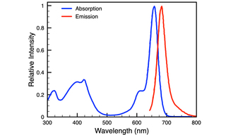
Figure 1. BeRST dye spectra.
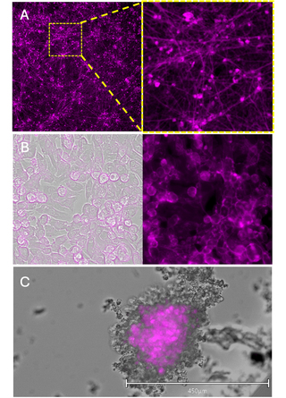
Figure 2. Cells labeled with BeRST dye. hIPSC-sensory neurons (A), hIPSC-cardiomyocytes (B), and 3D hIPSC-cardiomyocytes (C) loaded with1 µM BeRST dye. hIPSC-SN, 2D hIPSC-CM, and 3D hIPSC-CM were imaged at 10x, 20x, and 10x magnification, respectively. Scale bar only applies to C.
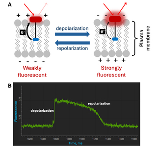
Figure 3. Voltage sensing with BeRST dye. (A) BeRST is a Photo-induced electron Transfer (PeT)-based voltage-sensitive dye that depends on the electron transfer from an electron-rich donor (blue) to the fluorophore (red) through a molecular wire (black). (B) Kinetic fluorescence waveforms from hIPSC-cardiomyocytes collected on the VOLTA Scanner demonstrating changes in transmembrane action potential voltage detected by monitoring BeRST dye fluorescence over time.
- Apr 16, 2025
- [1] YL Huang, AS Walker, EW Miller (2015) J Am Chem Soc 137:10767–10776(opens in new window)
- [2] A Klimas, G Ortiz, E Entcheva et al. (2020) Prog Biophys Mol Biol 154:62-70(opens in new window)
- [3] P Killfoil, SL Feng, S Jenkinson et al. (2021) Eur J Pharmacol 912:174584(opens in new window)
- [4] J Streit, S Kleinlogel (2018) Sci Rep 8:1153(opens in new window)
- [5] VOLTA Scanner: High-Throughput Cardiomyocyte Electrophysiology with an Optical Pacemaker (Lumencor Insight)